Fig. 3.
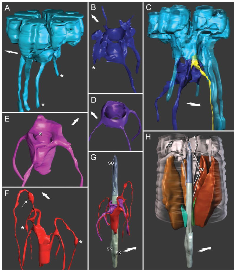
Three-dimensional reconstructions based on serial TEM sections of tissues and cuticle of the stomatostylet apparatus of Aphelenchus avenae. Body wall cuticle and all somal extensions are truncated posteriorly; body wall cuticle and most anterior epidermis (i.e. HypC) are truncated anteriorly through the tips of the lips. Large arrows indicate dorsal. Key to cells (given in Fig. 2). A: Lateral view of HypC toroid. Asterisks mark pseudosomal extensions. B: Anterior, ventral view of HypB toroid. Asterisk marks asymmetrically “missing” somal extension. C: Dorsolateral view of HypC (rendered semitransparent) and HypB toroids, showing the position of a representative inner labial socket cell. D: Anterior, ventral view of HypA toroid. E: Anterior, ventral view of toroid of anterior arcade syncytium. Asterisk indicates fold accommodating the tip of the vestibule extension. F: Ventrolateral view of toroid of posterior arcade syncytium. Thin arrow indicates swelling of somal extension at its contact point with dorsal somal extension of HypE and splitting somal extensions of HypC; asterisks indicate swellings of somal extensions at their contact points with the tips of the pseudosomal extensions of HypC. G: Ventrolateral view of stylet lined by arcade syncytia. Stylet cone is rendered transparent; stylet shaft material is shown projecting into thin wall (not lumen) of cone. Reconstruction of stylet shaft and knobs is a composite, the posterior portion (including knobs) reconstructed from a separate dataset of serial TEM sections. sk, stylet knobs (right and left subventral); so, stylet orifice. H: Ventrolateral view of the stylet with dorsal and left subventral stylet protractor muscles, as well as ventral and left subdorsal marginal cells. Cephalic framework (cf), to which protractors attach, guiding apparatus (i.e., vestibule, v, and vestibule extension, ve), and peripheral body wall cuticle are rendered transparent. Muscles and marginal cells are cut away anterior to their point of attachment to stegostom; right subdorsal marginal cell is hidden from view behind stylet; right subventral protractor has been removed for clarity.

