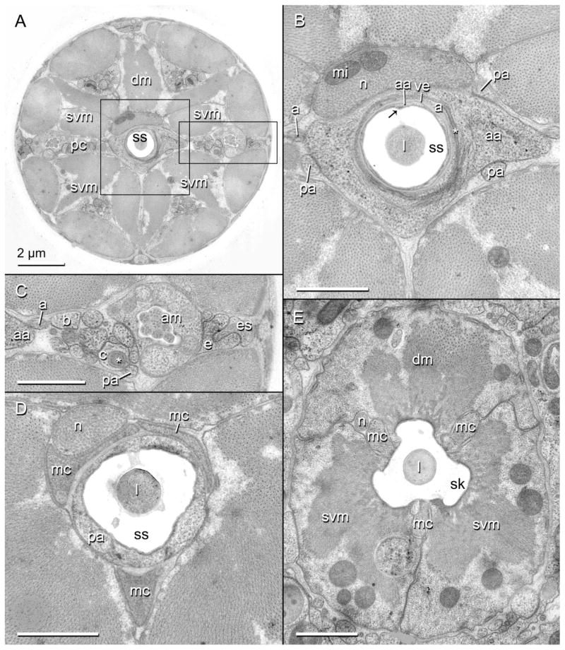
Fig. 5.
Transverse TEM sections through the gymnostom and stegostom of the head region of Aphelenchus avenae. Scale bars are 1 μm unless otherwise indicated. A: Entire transverse section of nematode showing the stylet shaft (ss), surrounding syncytia, and six robust pseudocoelomic cords (pc) separated by six incipient branches of the dorsal (dm) and subventral (svm) stylet protractor muscles. Area within central rectangle is enlarged in (B); area within rectangle to right is enlarged in (C). B: Enlarged inset of (A). Toroid of anterior arcade syncytium (aa) lining the stylet shaft and the thinned posterior tip of the vestibule extension (ve); asterisk marks tight junction between the anterior arcade cell and HypA (a); arrow indicates dorsal “seam” of stylet shaft. Also shown is the swollen tip of the pharyngeal neuron (n) associated with the dorsal protractor, including mitochondria (mi). l, stylet lumen; pa, posterior arcade syncytium. C: Enlarged inset of (A). A lateral pseudocoelomic cord, showing several syncytial extensions as identified by 3D reconstruction. Asterisk marks one of the multilamellar dendrites just anterior to its entry into the pseudosomal extension of HypC (c). am, amphid; b, HypB; e, HypE; es, mid body epidermal syncytium. D: Toroid of posterior arcade syncytium lining the posterior part of the stylet shaft and anterior extensions of the marginal cells (mc). E: Triradiate stylet protractor muscles and marginal cells lining the stylet knobs (sk).

