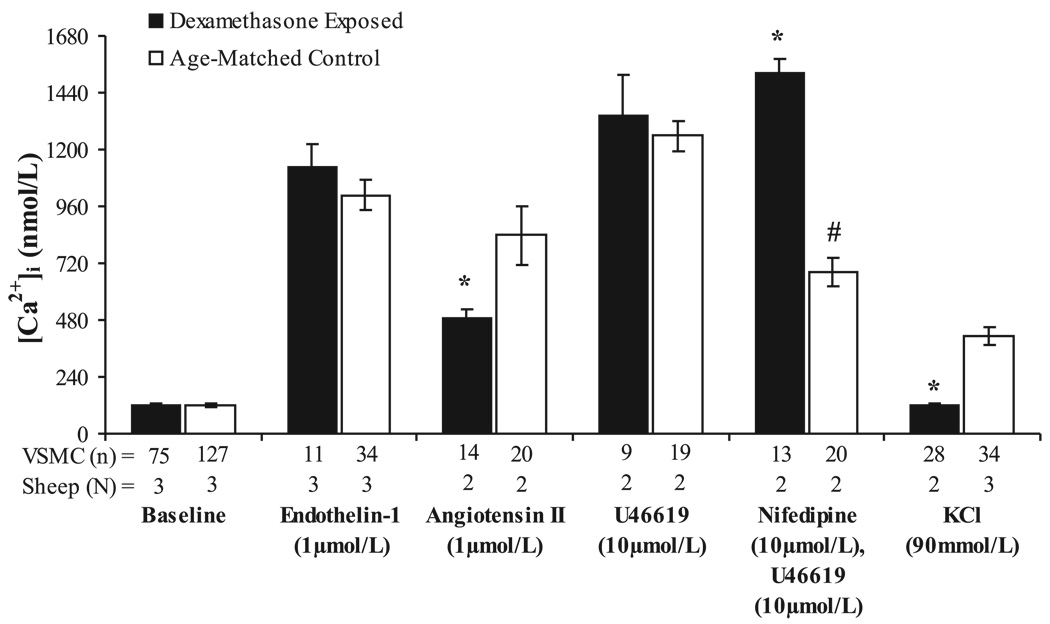
Fig. 3.
Vascular smooth muscle cell cytosolic calcium concentration measured at baseline and following stimulation with endothelin-1, ANG II, U-46619 with and without nifedipine pretreatment, or KCl in dexamethasone-treated and control coronary artery cells. Values are displayed as means with vertical lines indicating SE; N = number of lambs (and coverslips); n = number of VSMC analyzed. *Significant differences between dexamethasone-exposed and control lambs (P < 0.05). #Significantly different than response of control VSMC to U-46619 alone (P < 0.01).

