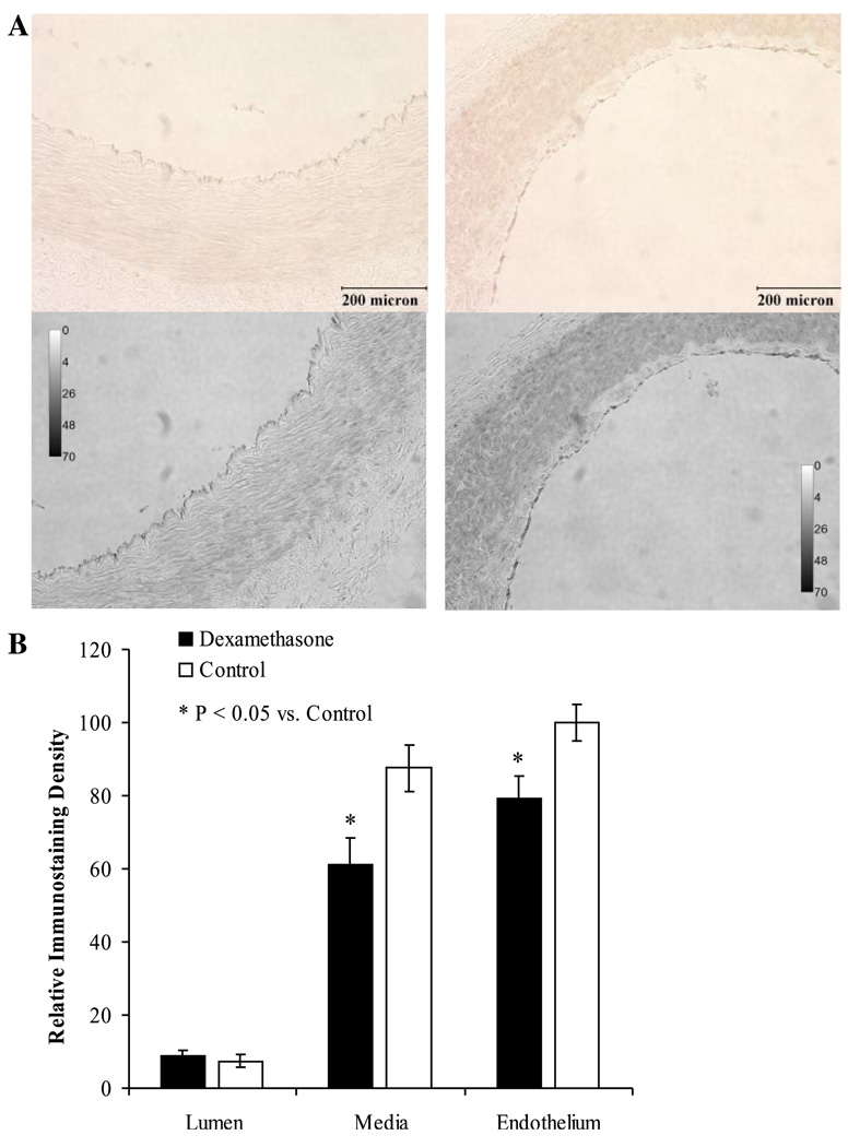
Fig. 5.
Immunostaining for eNOS protein within coronary arteries harvested from dexamethasone-exposed (left) and control (right) newborn lambs (A). The first row demonstrates the brown immunostaining seen using the peroxidase substrate diaminobenzidine. The images were then converted to gray scale (A, second row) before quantification by densitometric analysis (B). Relative immunostaining density (as a reflection of eNOS expression) was significantly localized to the vascular endothelium (P < 0.01 vs. the media for each treatment group) and was significantly attenuated following early gestation dexamethasone-exposure. *Significantly different from control (P < 0.01). Values are displayed as means with vertical lines indicating SE and magnification ×200 was used for each image.

