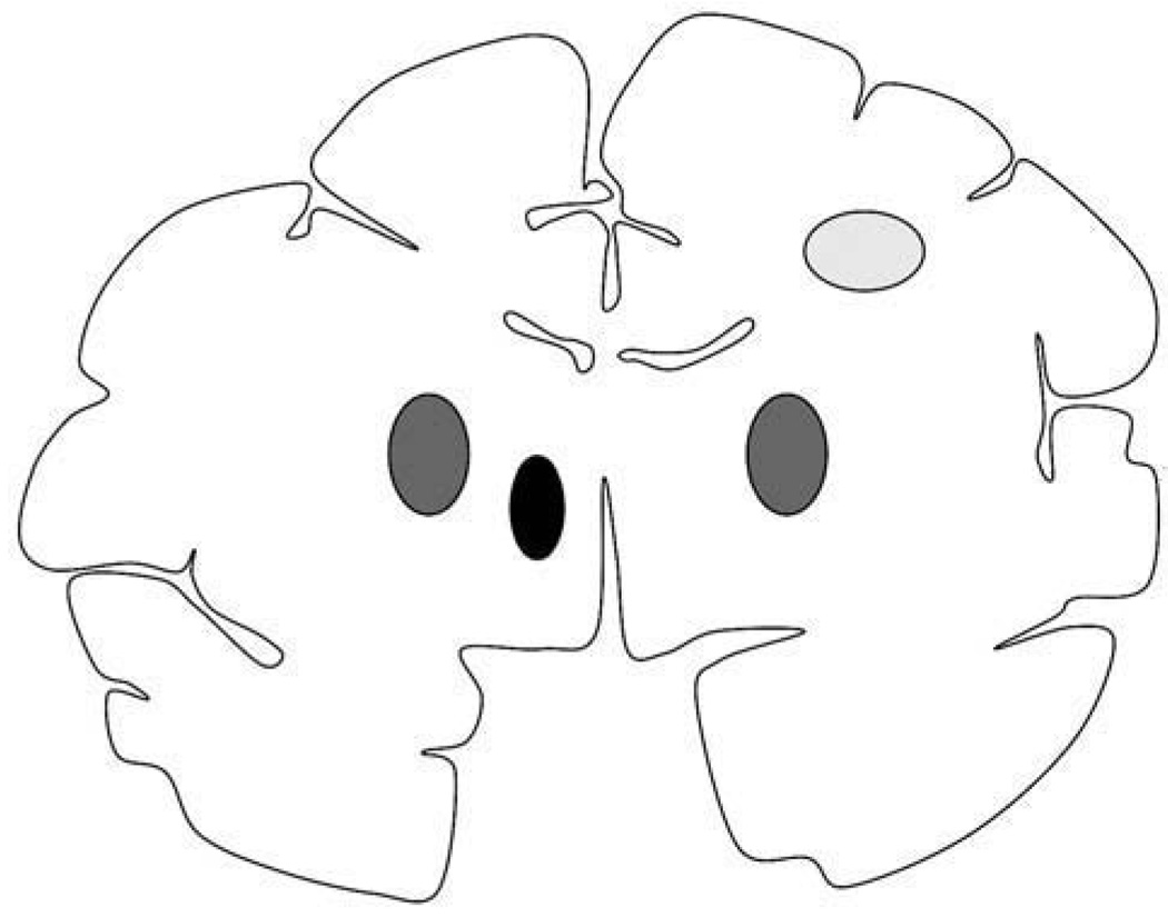
Fig. 1.
Schematic representation of the non-human primate brain in coronal section and sites of infusion. Region of corona radiata infused (light gray oval) in the right hemisphere in all three animals (NHP1, NHP2, and NHP3). Bilateral putaminal convections (dark gray ovals) were present in two NHP, whereas the remaining animal had only putaminal infusion in the right hemisphere. All three animals received thalamic infusion (black oval) in the left hemisphere.

