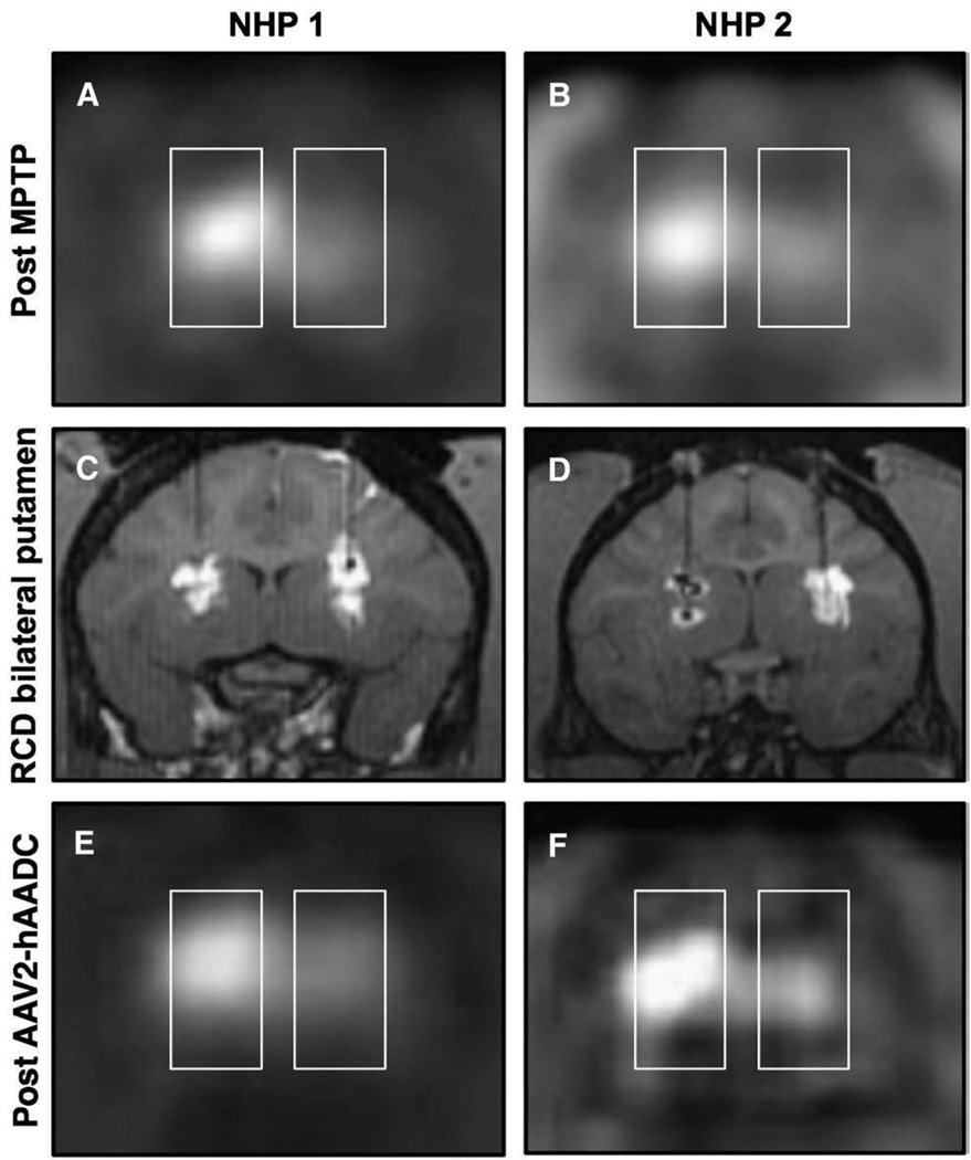
Fig. 3.
Coronal FMT-PET images after intracarotid MPTP. Panels A and B show a clear reduction in PET signal on the side ipsilateral to the intracarotid infusion. Bilateral RCD of putaminal targets (C and D) reveal robust bilateral GDL distribution in one subject (C), and MRI evidence of oil/air along with convection within the left putamen and otherwise robust convection within the right putamen of the other subject (D). Repeat FMT-PET after AAV2-hAADC gene therapy (E and F) shows a mild increase in bilateral PET signal in one subject (E), and marked increase in bilateral PET signal in the other animal (F).

