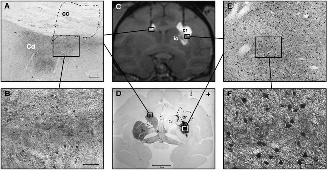
Fig. 6.
MRI correlation with histology in NHP with bilateral attempted putaminal AAV2-hAADC RCD. Panel A shows a low magnification view of the dorsolateral left head of the caudate (Cd) and overlying corpus callosum (cc). AADC staining is seen within the Cd after a medially placed cannula tract allowed it to be infused into this region rather than the left putamen. Notice the lack of specific AADC staining in the overlying corpus callosum (dotted polygon), despite GDL signal in this region on MRI (see C). Scale-bar=0.25 mm. Panel B depicts a representative view of higher magnification AADC staining in the left head of caudate. No such cellular staining was noted in the overlying white matter tracts. Scale-bar=0.10 mm. Panel C displays a coronal T1-weighted MR image of NHP brain with the bilateral RCD with GDL contrast. Although the putaminal targeting was better in the right hemisphere, there was some spread of the GDL into the internal capsule (ic) and overlying corona radiata (cr). The left-sided targeting was too medial and allowed convection of the head of the caudate and the overlying corpus callosum. Panel D shows a coronal histologic brain section of the same animal as imaged in panel C. This section is double-labeled, showing TH immunoreactivity (TH-IR) within the left striatum and paucity of TH-IR on the right, as a result of the right intracarotid MPTP infusion. The dense immunoreactivity in the left head of the caudate represents AADC IHC staining. Similar but more abundant AADC staining is seen within the substance of the right putamen, and part of the head of the right caudate. Note the absence of AADC staining within the overlying right corona radiata (dashed polygon, cr) and internal capsule, despite both of these regions showing GDL enhancement in panel C. Panel E depicts a low magnification view of the boxed region within the right putamen shown in D. Note the specific neuronal staining for AADC. Scale-bar=0.25 mm. Panel F shows a higher magnification view of the boxed region depicted within E. Notice the specific staining of medium spiny neurons. Scale-bar=0.10 mm.

