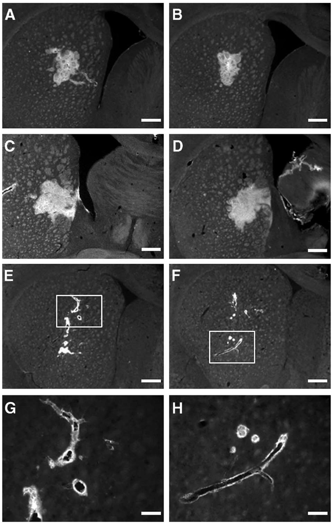
FIG. 2.
Photographs of brain sections stained for AAV2 capsids (antibody A20) in rats after intrastriatal infusion of AAV2 by convection-enhanced delivery. (A and B) Striatal sections from two different rats from group H (high blood pressure/heart rate); (C and D) brain sections from a more caudal part of the same brains showing positive signal in globus pallidus. For all rats from group H, the fluorescent signal was spread evenly within the hemisphere, forming a mostly uninterrupted distribution. (E and F) Brain sections from two rats with low blood pressure/heart rate when AAV2 was infused. The signal is restricted to the vicinity of infusion and the perivascular area surrounding nearby blood vessels. (G and H) Higher magnification views of E and F (original magnification ×20). Size bars: 500 (A through F) and 125 µm (G and H).

