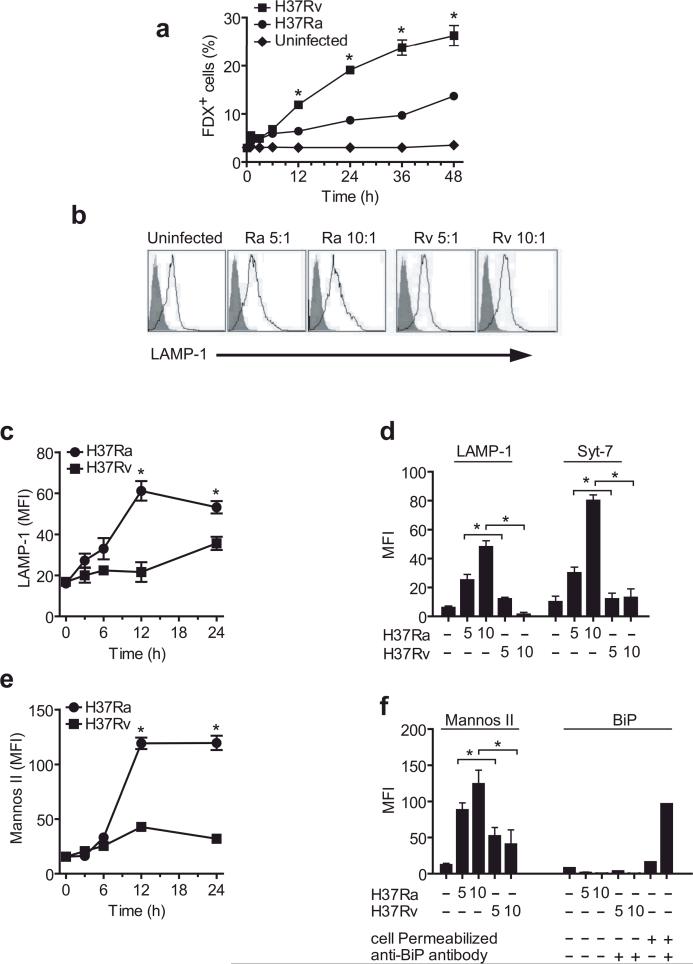
Figure 1. Infection of human Mφ with virulent H37Rv inhibits lysosomal and Golgi-mediated plasma membrane repair.
(a) Kinetics of FDX influx through membrane lesions in Mφ left uninfected or infected with H37Ra or H37Rv. (b) LAMP1 translocation to the plasma membrane lesions of human Mφ infected with H37Ra or H37Rv for 12 h. Shaded histogram is the isotype control and the open histogram is the specific LAMP1 staining. Numbers in histograms indicate the mean fluorescence intensity (MFI) of the entire cell population. Bacterial strain and MOI is indicated above each histogram. (c,e) Kinetics of LAMP1 (c) and mannosidase II (e) translocation to the surface of human Mφ infected with H37Ra or H37Rv. (d, f) Translocation of LAMP1, Syt-7, mannosidase II and the ER marker BiP to the surface of Mφ left uninfected or infected for 12 h with H37Ra or H37R. Where indicated, Mφ were permeabilized and stained with irrelevant (−) and BiP-specific (+) antibodies. The MOI was 5:1 (5) or 10:1 (10) where indicated; otherwise an MOI of 10:1 was used. Results in all panels are representative of at least three independent experiments (error bars, s.e.m.) *, p < 0.05.

