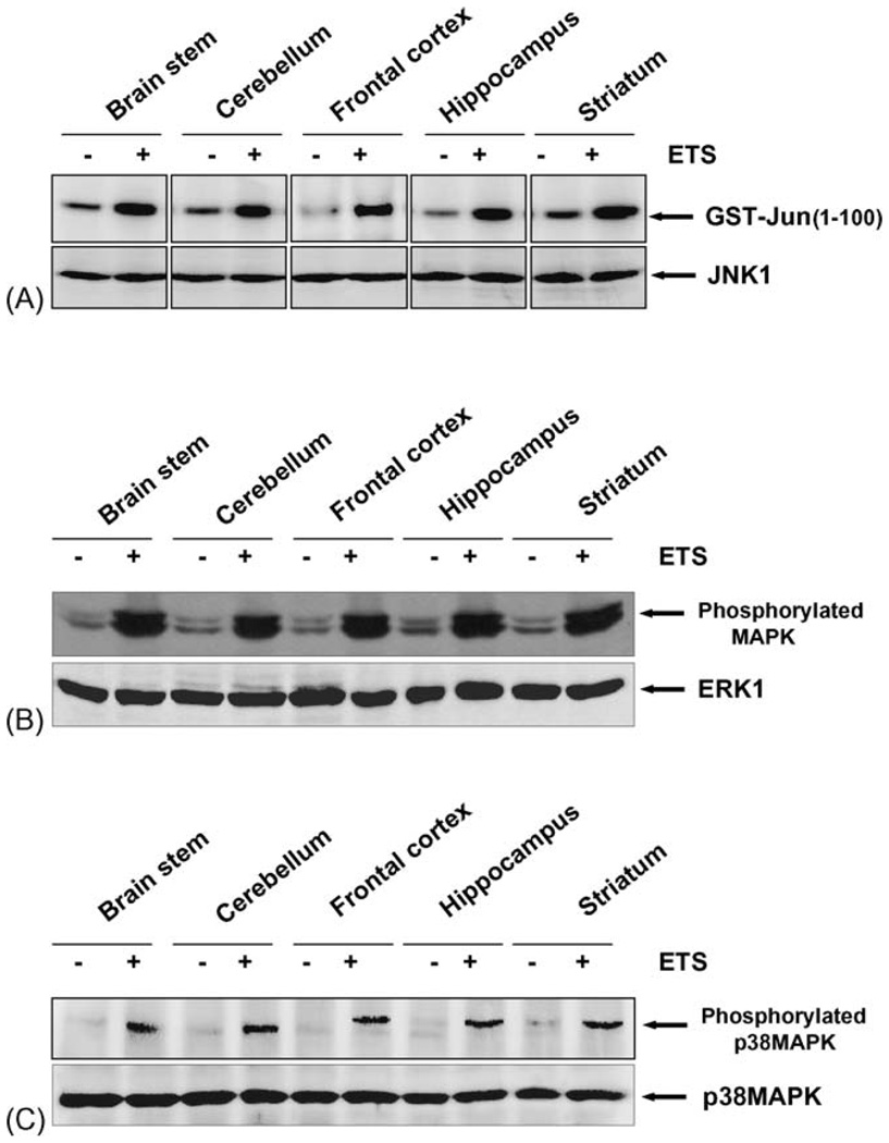
Fig. 4.
(A) ETS induces JNK activation. Tissue extracts from different regions of brain were collected and 300 µg proteins were immunoprecipitated with anti-JNK (0.3 µg/sample) antibody, and the kinase reaction was performed using 32P-labeled ATP and GST-Jun as substrate protein. The radiolabeled GST-Jun was detected in 9% SDS-PAGE. The level of JNK was detected using 50 µg of same extract proteins and analyzed in 10% SDS-PAGE by Western blot. (B) ETS induces activation of MAPK. Tissue extracts from different regions of brain were collected, (100 µg) of the protein was analyzed by 10% SDS-PAGE and detected phosphorylated MAPK using anti-phospho MAPK antibody by Western blot. The same blot was stripped off and re-probed with anti-ERK1 antibody to detect the ERK1 by Western blot. (C) ETS induces activation of p38MAPK. Tissue extracts from different regions of brain were collected, (100 µg) of the protein was analyzed by 10% SDS-PAGE and detected phosphorylated p38MAPK using anti-phospho p38MAPK antibody by Western blot. The same blot was stripped off and re-probed with anti-p38MAPK antibody to detect the levels of p38MAPK by Western blot.

