Fig. 2.
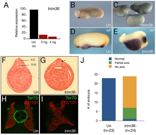
Depletion of maternal trim36. (A) Quantitative real-time PCR of trim36 expression in oligo-injected oocytes. Samples were normalized to odc levels and displayed as a percentage relative to the uninjected oocyte (Un oo) sample. Oocytes were injected with 3.0 or 4.0 ng of trim36-3 oligo. (B) Phenotype of an uninjected stage 24 embryo. (C) Phenotypes of sibling trim36-depleted embryos. (D) In situ hybridization of sizzled (szl) in an uninjected control embryo. (E) szl expression in a trim36-depleted embyro. (F) H&E-stained section of a control embryo, with the neural tube (n.t.) and notochord (n.o.) indicated. (G) H&E-stained section of a trim36-depleted embryo; notochord and neural tube are absent, and a somite muscle mass persists in the midline (arrow). (H,I) Notochord marker Tor70 (green) and somite marker 12/101 (red) in control (H) and trim36-depleted (I) embryos. (J) Histogram showing the distribution of phenotypes (see key) in trim36-depleted embryos (from two experiments).

