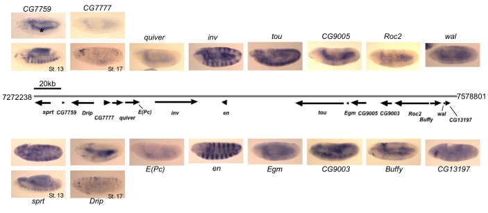
Fig. 1.
Expression patterns of en, inv and flanking genes in Drosophila embryos. Gray line indicates genomic DNA, with the coordinates listed at both ends (genome version R5.1, FlyBase). Arrows below indicate the transcription units and the direction of transcription. In situ hybridization was performed on embryos using DIG-labeled RNA antisense probes and alkaline phosphatase staining. All embryos are stage 11 unless otherwise noted. For some genes, an additional image of a later stage embryo is shown to demonstrate a specific staining pattern if no pattern was observed at stage 11. Several genes show weak ubiquitous expression or do not show specific staining at any stage in embryos. Non-specific yolk sac staining is observed in many embryos, indicated with an asterisk in CG7759. All embryos are oriented anterior to the left and dorsal up.

