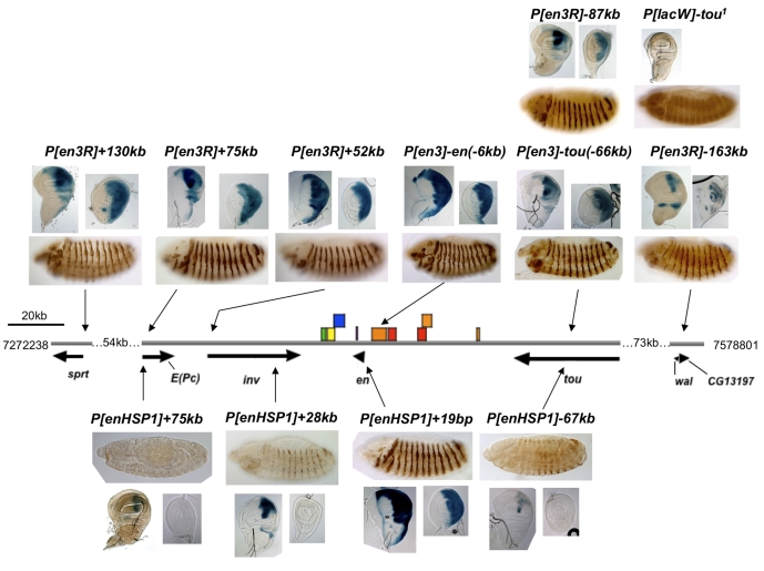
Fig. 3.
Expression patterns of enhancer traps in the en/inv genomic region. Gray line indicates genomic DNA. Arrows under the genomic DNA line show the position and direction of transcription units. Vertical arrows indicate the position of insertion of the transgene indicated. The exact insertion site of each P-construct is listed in Table 1. Transgenes with the en promoter are above the genomic DNA line, those with the heat shock promoter are below the genomic DNA line. The position of all known en stripe enhancers and other enhancers important for these experiments are shown as colored boxes: green, clypeolabrum; yellow, posterior spiracles; blue, hindgut; orange, stripe in every segment; red, even or odd stripes (pair-rule enhancers); purple, en intron enhancer: hindgut, posterior spiracles, fat body, even stripes and every segment (Kassis, 1990) (our unpublished data). The imaginal disc enhancer is upstream of en but the exact location is not known (unpublished data). β-Galactosidase protein is visualized by immunoperoxidase staining in embryos and X-gal staining in imaginal discs. A lateral view of a germ-band-shortened embryo (stage 13), ∼10 hours after egg laying, with anterior to the right and dorsal up, is shown for each transgene. A wing (left) and leg (right) imaginal disc (posterior compartment to the right) is also shown for each transgene (except P[lacW]-tou). Discs were fixed in formaldehyde and stained for varying amounts of time to equalize staining intensity. Lines with P[en3] or P[en3R] insertions in en or inv stained within 30 minutes; E(Pc),1 hour; tou, 2.5 hours; sprt and wal, 5 hours. Note that the expression in the posterior compartment of the P[en3R]-131 line is variegated, suggesting that activation by en enhancers in some cells is stronger than in others. This variegation is not evident when the discs are stained for a longer period of time. For imaginal disc staining, lines P[enHSP1]+28kb, +19bp and -67kb were fixed with formaldehyde and stained for varying times: P[enHSP1]+19bp, 30 minutes; P[enHSP1]+28kb, 2.5 hours; P[enHSP1]-67kb, 48 hours. Line P[enHSP1]+75kb was fixed with glutaraldehyde and stained for >24 hours. We were not able to see β-gal activity in this line when it was fixed with formaldehyde.

