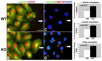
Fig. 7.
Defect in the specification of cell polarity in Cx43 KO epicardial cells. (A-D) Epicardial cells from E11.5 explant cultures were immunostained with antibodies to α-tubulin (A,B), or γ-tubulin and the Golgi GM130 marker (C,D) to visualize the MTOC. α-Tubulin staining exhibited an intensely stained crescent-shaped structure that corresponds to the MTOC. In wild-type cells, most of the MTOC are forward facing, i.e. situated in the leading edge of the cell (white arrow indicates the direction of cell migration). The microtubules were also observed to align with the direction of cell migration. By contrast, in the Cx43 KO explants, the MTOC and the microtubule cytoskeleton were often not aligned with the direction of cell migration (B). A similar result was obtained when γ-tubulin and GM130 antibodies were used to track the positioning of the MTOC in wild-type (C) and KO epicardial cells (D). The quantitative analysis is shown in the bar graphs to the right. Red and blue staining in A,B and C,D, respectively, corresponds to DAPI staining. Scale bar: 50 μm.

