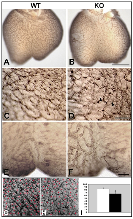
Fig. 9.
Defect in remodeling of the primitive coronary vascular plexus in Cx43 KO mice. (A-H) Whole-mount Pecam immunostaining of E13.5 hearts shows the distribution of the coronary vascular plexuses in wild-type (A,C,E,G; WT) and Cx43 KO (B,D,F,H; KO) mouse hearts. (A-D) Wild-type (A,C) and Cx43 KO (B,D) mouse hearts show a network of coronary vascular plexuses. In enlarged views, the KO (D), but not the wild-type (C) heart shows fine vessels (arrowheads) criss-crossing the major coronary vascular network. (F) Magnified anterior view of Pecam-stained Cx43 KO (F) heart shows abnormally enlarged vessels near the apex. (E) Similar view of a Pecam-stained heart from a wild-type littermate. (G,H) Enlarged images of the hearts in A and B with red dots showing the scoring of vascular network. The results of this analysis, plotted in I, show a reduction in the density of the coronary vascular network in the KO (H, black bar in I) when compared wih wild-type (G, white bar in I) hearts. Scale bars: 50 μm in B (same as A); 10 μm in D and F (C,D and E,F are same magnification).

