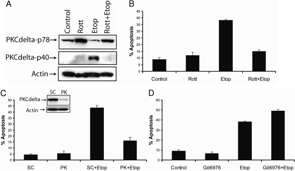
Fig. 2.
Etoposide induces PKCδ-mediated apoptosis in SK-N-AS cells. A, immunoblot analysis of PKCδ and β-actin in cell lysates after the treatment with 2 μM rottlerin, 50 μM etoposide, or 2 μM rottlerin and 50 μM etoposide for 48 h. B, cells were treated as described above, and the percentage of apoptotic cells was determined by staining with annexin V and propidium iodide and analyzed by flow cytometry. All values are representative of three independent experiments, and error bars show S.D. from triplicate measurements. C, immunoblot analysis of PKCδ and β-actin from whole-cell lysates after the transfection with 100 nM nontargeting siRNA (SC) or PKCδ -specific siRNA for 72 h (inset), cells were treated with 50 μM etoposide for an additional 48 h, and apoptosis was determined by counting fragmented nuclei after staining with Hoechst 33342 among 200 cells. Shown are representative apoptosis rates from three independent counts. D, cells were treated with 10 nM Gö6976 for 2 h followed by 50 μM etoposide for 48 h, and the percentage of apoptotic cells was determined by staining with annexin V and propidium iodide and analyzed by flow cytometry. All values are representative of three independent experiments, and error bars show S.D. from triplicate measurements.

