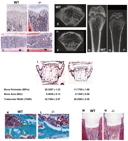
Fig. 4.
Gpr48 regulates bone formation postnatally. (A-D) H&E staining of tibia of wild-type (A,C) (10×) and Gpr48-/- (B,D) (10×) mice at P18. There is much less trabecular bone in Gpr48-/- than wild-type mice, and the cortical bone is much thinner in the mutant tibia (arrows). (E-H) Transverse (E,F) and frontal (G,H) microCT sections of distal and midfemoral diaphyses of 1-month-old wild-type and Gpr48-/- mice. (I,J) von Kossa staining of L3 vertebral bodies from 1-month-old wild-type and Gpr48-/- mice. There is a significant reduction in bone perimeter, bone area and trabecular width in Gpr48-/- mice. (K,L) Osteoid synthesis defect in Gpr48-/- mice demonstrated with Goldner staining (osteoid is stained red, yellow arrows). Deletion of Gpr48 significantly reduced osteoid formation (compare K with L). (M,N) Masson-Trichrome staining of osteoid (blue).

