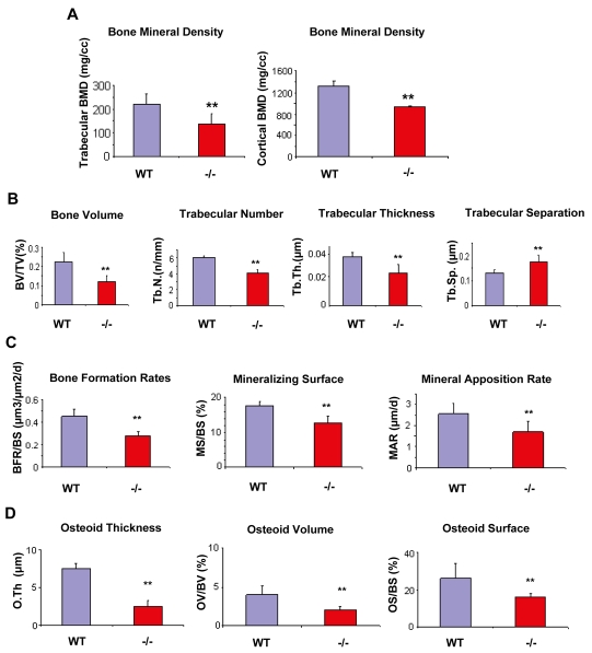
Fig. 5.
Quantitative analysis of the osteoblast defect in Gpr48-/- mice. (A) Both trabecular bone mineral density (BMD) and cortical BMD dramatically decreased in Gpr48-/- mice as assessed on microCT sections. (B) Deletion of Gpr48 leads to a reduction in bone volume/tissue volume (BV/TV), trabecular thickness, trabecular number, and increased trabecular separation as assessed by histomorphometric analysis. (C) Gpr48 regulates the kinetic indices of mineral apposition (MAR), bone formation rates (BFR/BS) and mineralizing surface (MS/BS). (D) Deletion of Gpr48 causes significant defects in osteoid characteristics, including decreased osteoid thickness (O.Th), osteoid surface (OS/BS) and osteoid volume (OV/BV). In each case, results show mean±s.d., n=3, age and sex matched; **P<0.01 in A-D.

