Fig. 5.
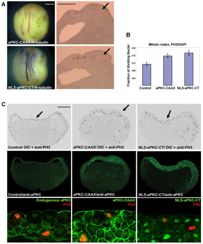
Effect of overexpressing aPKC constructs on cell proliferation. (A) Neurula stage Xenopus embryos overexpressing aPKC-CAAX and NLS-aPKC-CT showed suppression of N-tubulin expression and a thickened ectoderm (arrows). (B) Quantitation of phospho-histone H3 (PH3)-positive nuclei relative to the total number of nuclei as obtained from at least three embryos in each set and a minimum of five sections per embryo. (C) Sections of st.10.5 embryos injected with aPKC-CAAX and NLS-aPKC-CT and co-immunostained with anti-PH3 (black in DIC image, top row, and red in bottom row) and anti-aPKC (green, in middle and bottom rows) antibodies. Sections show multilayered, thickened ectoderm and increased PH3 staining in both layers of the ectoderm. The bottom row shows the middle row at higher magnification, with anti-aPKC and anti-PH3. Scale bars: 250 μm in A; 50 μm in C.

