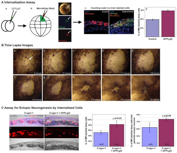
Fig. 8.
Lgl2-expressing cells become internalised and contribute to primary neurogenesis. (A) The distribution of GFPLgl2-expressing cells in the inner versus outer ectodermal layer of neurula stage Xenopus embryos was assessed by an internalisation assay (see Materials and methods for details) that involved lineage-labelling individual GFPLgl2-expressing outer cells by Micro-Ruby injection at the blastula stage (a,b). The fraction of Micro-Ruby-labelled cells in the inner layer over the total number of labelled cells was increased in GFPLgl2-injected embryos as compared with control embryos (c,d). (B) Time-lapse images of an embryo overexpressing GFPLgl2, showing early loss of apicobasal cell polarity in superficial cells (apical de-pigmentation, arrow in a) and subsequent internalisation of these cells at mid-gastrula stage (g-j). (C) To determine whether internalised cells resulting from GFPLgl2 overexpression contribute to increased neurogenesis, the Micro-Ruby-based internalisation assay was combined with in situ hybridisation for N-tubulin, as analysed in sections of st.16 neurulae from embryos that had been injected at the 2-cell stage with either X-ngnr-1 alone or in combination with GFPLgl2 (a-f). Bar charts show that the percentage of Micro-Ruby-labelled deep layer cells relative to the total number of labelled cells was increased when GFPLgl2 was co-injected and that ∼76% of these cells, on average, were also N-tubulin positive. Scale bars: 500 μm in B; 50 μm in C.

