Fig. 1.
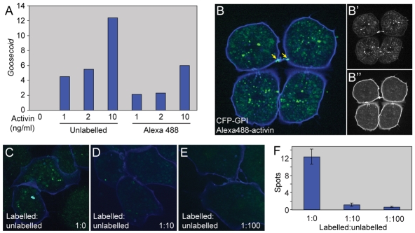
Alexa488-activin internalisation in dissociated animal cap cells. (A) Real-time RT-PCR analysis of dissected animal pole tissue treated with the indicated amounts of unlabelled activin or Alexa488-activin and cultured to the equivalent of the early gastrula stage. Bars indicate induction of Gsc. Note that unlabelled activin is approximately twice as effective as Alexa488-activin in inducing Gsc. This experiment was carried out four times, with similar results each time. (B-B″) Dissociated animal pole cells at the late blastula stage following incubation in Alexa488-activin for 1 hour before washing. Cells were derived from embryos that had previously been injected with the membrane marker CFP-GPI (blue). (B′) Alexa448 fluorescence alone. (B″) CFP-GPI fluorescence alone. Note Alexa488-activin accumulation at a cell-cell bridge (arrow in B). (C-F) Competition experiment in which cells are treated with Alexa488-activin in the presence of increasing amounts of unlabelled activin. (C) Internalisation of Alexa488-activin in the absence of unlabelled activin. (D) Internalisation of Alexa488-activin in the presence of a 10-fold excess of unlabelled activin. (E) Internalisation of Alexa488-activin in the presence of a 100-fold excess of unlabelled activin. (F) Quantitation of the experiment illustrated in C-E, showing mean numbers of aggregates (spots) of internalised Alexa488-activin. Values are means of 20-30 cells.

