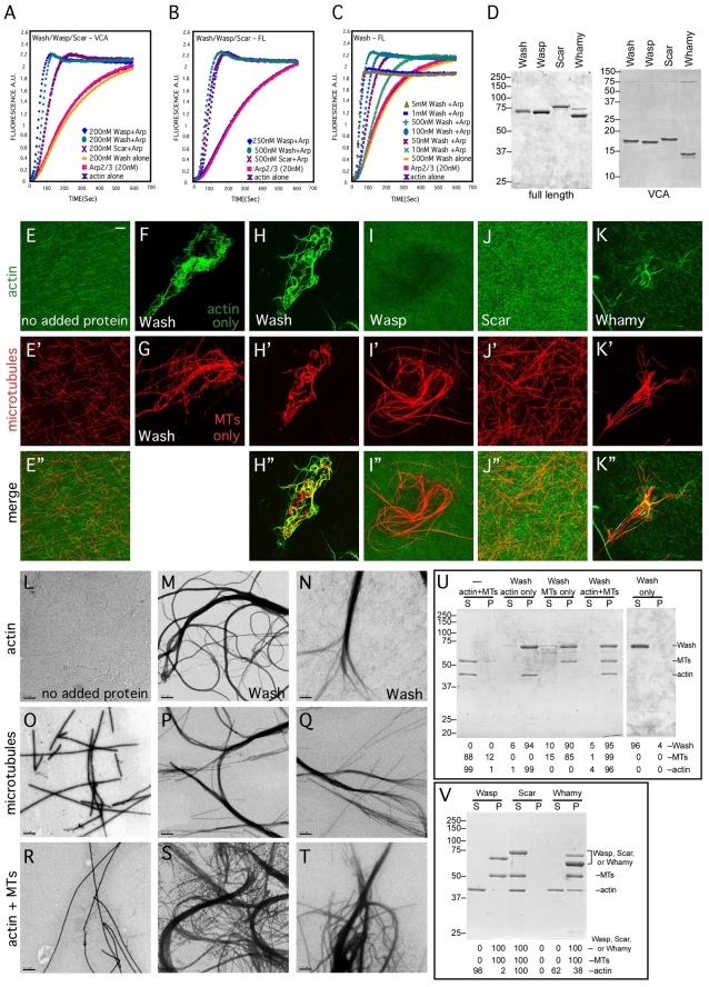
Fig. 1.
Wash activates Arp2/3 to nucleate actin, and bundles F-actin and microtubules. (A-C) Pyrene-actin polymerization assays. Wash, Wasp and Scar VCA fragments (A) and full-length (FL) proteins (B) nucleate actin in the presence of Arp2/3. Wash nucleation activity is concentration dependent and requires Arp2/3 (C). All polymerization assays were performed a minimum of three times. (D) Coomassie-stained protein gels of full-length and VCA fragments of Wasp family proteins used in this study. (E-K″) Stabilized F-actin (1 μM; top) and MTs (1 μM; middle) were incubated with WAS family proteins and observed by confocal microscopy: (E) no protein added; (F) Wash with actin only; (G) Wash with MTs only; (H-H″) Wash; (I-I″) Wasp; (J-J″) Scar; (K-K″) Whamy. (L-T) Electron micrographs of negatively stained samples from the indicated F-actin and MT bundling/crosslinking assays above. Two examples are shown for each assay with Wash (M-T). (U,V) Quantification of F-actin and MT bundling/crosslinking efficiency of indicated reactions by low-speed pelleting assays. The crosslinking/bundling reactions described above were centrifuged to pellet actin and microtubule bundles. Supernatants (S) and pellets (P) were separated and analyzed by Coomassie staining. Percentages of protein, actin and microtubules in each fraction are given. All assays were performed a minimum of three times. Final protein concentrations: Wash, 300 nM; Wasp, 300 nM; Scar, 300 nM; Whamy, 300 nM. Scale bars: 10 μm for E-K″; 500 nm for L-T.

