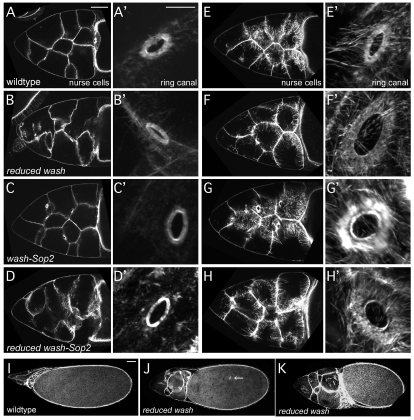
Fig. 6.
Wash functions with Arp2/3 to regulate actin cyto-architecture in oogenesis. (A-H′) Nurse cells (A-H) and ring canals (A′-H′) from stage 10a (A-D′) and stage 10b-11 (E-H′) egg chambers fixed and stained with phalloidin. Reduced wash (wash/+; wimp/+ females), wash-Sop2 (Sop2 wash/+ + females) and reduced wash-Sop2 (Sop2 wash/+ +; wimp/+ females) mutant oocytes (B-D,F-H) show a loss of nurse cell integrity and incomplete stress fiber formation compared with wild type (A,E). Reduced wash, wash-Sop2 and reduced wash-Sop2 mutant oocytes do not affect ring canal formation (B′-D′), but do exhibit abnormal actin organization around ring canals by stage 10b-11 (F′-H′). (I-K) Stage 14 egg chambers fixed and stained with phalloidin. Compared with wild type (I), reduced wash mutant nurse cells (J,K) retain more of their cytoplasm (incomplete dumping), and oocytes are smaller. Consistent with defects in actin cyto-architecture, ring canals separated from the nurse cell membrane can be found `floating' within the oocyte (arrow in H). Scale bars: in A, 50 μm for A-H; in A′, 10 μm for A'-H'; in I, 50 μm for I-K.

