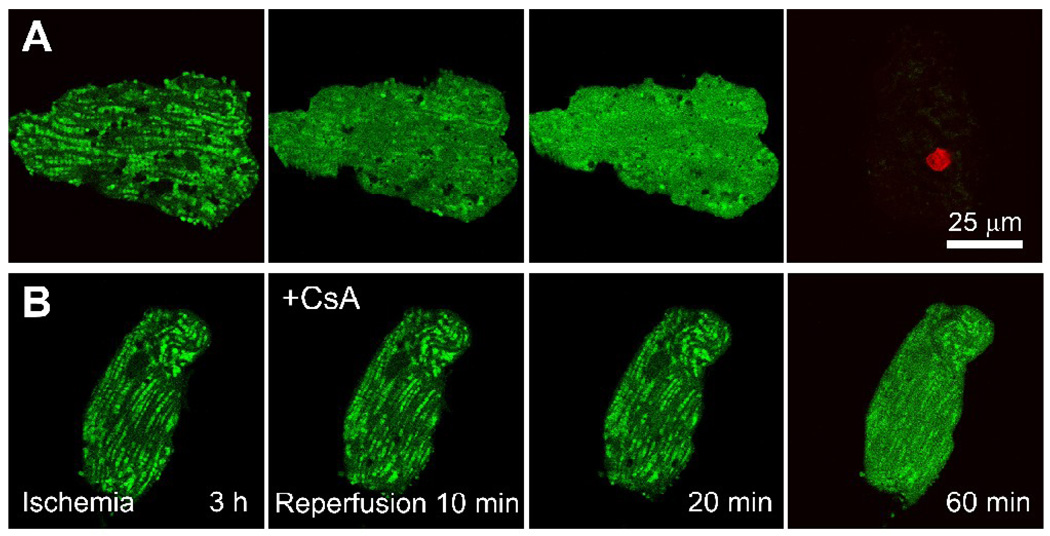
Figure 2. The MPT and cell death after ischemia-reperfusion to adult rat cardiac myocytes.
Mitochondria of myocytes were loaded with calcein by a cold-loading/warm incubation procedure, and the cells were subjected to 3 h of anoxia at pH 6.2 (ischemia) followed by reoxygenation at pH 7.4 (reperfusion) in the absence (A) and presence (B) of 1 µM CsA. Nuclear staining by red-fluorescing propidium iodide (PI) detected loss of cell viability. Note that green mitochondrial calcein fluorescence was retained at the end of ischemia, indicating that PT pores were closed. After reperfusion in the absence of CsA (A), mitochondria released calcein into the cytosol beginning after as early as 10 min. After 60 min, all cellular calcein was lost, and PI nuclear staining signified loss of cell viability. In the presence of CsA added at reperfusion (B), mitochondrial release of calcein was greatly diminished and viability was maintained for more than an hour. Adapted from [26].

