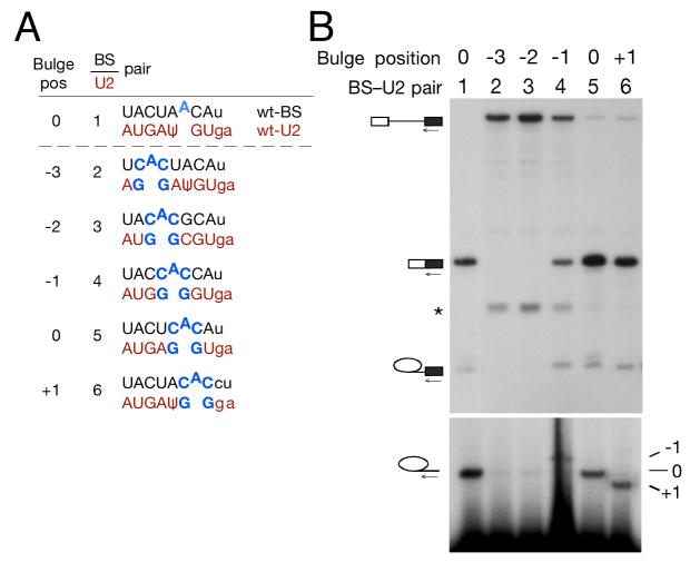
Figure 2. Systematic variation of bulge position indicates that at least three positions within the BS-U2 duplex are competent for first step catalysis.
(A) Schematic of reporter and U2 constructs used in (B).
(B) Primer extension with a 3′ exon primer indicates that −1, 0 and +1 BS bulge positions allow splicing catalysis. In this and subsequent figures, a band resulting from degradation of pre-mRNA is marked by an asterisk.

