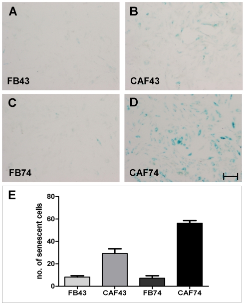
Figure 6. Senescence-associated beta-galactosidase activity of fibroblasts.
Representative pictures of fibroblast monolayer cultures stained for SA-β-gal activity. FB-43 (A) and FB-74 (C) show little or no staining; CAFs are positive (B and D). Scale bar 200 µm. CAF-74 had significantly more (P<0.01) SA-β-gal cells compared to CAF-43 (E). Columns: mean; error bars; SEM.

