
Figure 3.
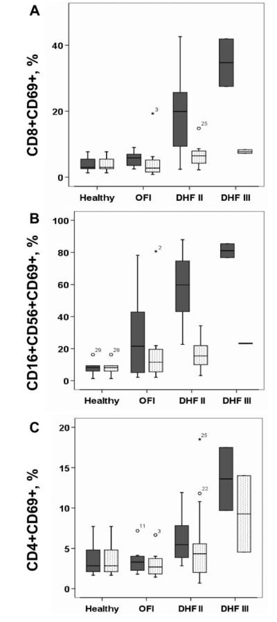
Cellular activation during acute dengue. Shown are the median, interquartile, and 95th-percentile percentage ranges of (A) CD8+CD69+ T cells, (B) CD3-CD16+CD56+CD69+ NK cells, and (C) CD4+CD69+ T cells, as a proportion of lymphocytes in peripheral blood from healthy infants (n = 6) and from infants who had either dengue hemorrhagic fever grade II (DHF II) (n = 15) or III (DHF III) (n = 2) or other febrile illness (OFI) (n = 8), at study enrollment (shaded boxes) and at discharge (unshaded boxes). At the time of study enrollment, infants with acute DHF II had significantly higher percentages of CD8+CD69+ T cells (P = .006, by Mann-Whitney test), CD16+CD56+CD69+ NK cells (P = .001, by Mann-Whitney test), and CD4+CD69+ T cells (P = .04, by Mann-Whitney test) than did infants with OFI.