
Figure 1.
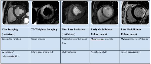
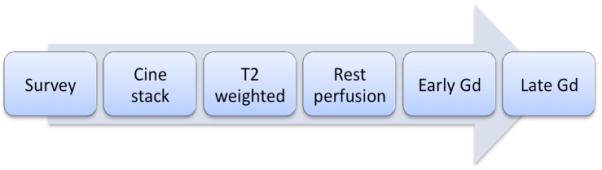
Figure 1a. CMR methods for assessment of ACS. The figure shows short axis views (of different patients) illustrating the different imaging techniques used, their morphological correlates main clinical application.
Figure 1b. These methods can be integrated into a suggested CMR protocol that provides a comprehensive assessment of ACS patients and can be performed in less than 1 hour. Gd = gadolinium.