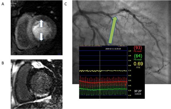
Figure 6.
CMR images from a patient with previous stents to the RCA and LAD 2 years earlier who presented to the emergency room with troponin-negative chest pain. CMR was performed within 24 hours of admission. Panel A shows inferior and anterior ischemia on adenosine-stress perfusion imaging. Panel B shows late gadolinium enhanced images with a region of subendocardial gadolinium enhancement in the inferior wall suggesting old myocardial infarction that was not previously known about. T2-weighted images were normal (not shown). Subsequent coronary angiography revealed tight in-stent restenosis in the RCA and significant flow-limiting disease in the LAD (panel C) as assessed by a pressure wire during hyperaemic conditions (fractional flow reserve 0.69). Both lesions were stented successfully and the patient was discharged home.

