
Figure 5. Phosphorylation of Histone H2AX (γ-H2AX) in BxPC3 and Panc1 cells following treatment with Gem ± PD-321852.
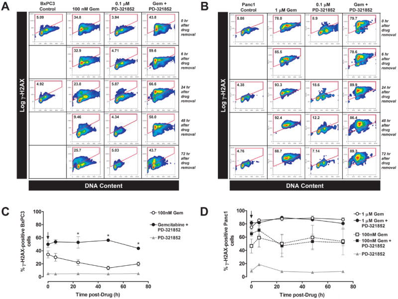
BxPC3 cells were treated with 100 nM Gem ± 0.1 μM PD-321852 for 24 h. The agents were then washed out (as indicated by the arrow in (C)), and cells were collected either immediately, or after incubation in control medium for an additional 6, 24, 48 or 72 h. Cells were analyzed by flow cytometry for the presence of γ-H2AX staining, as a function of DNA content (A & C). The number inside the gate is the percentage of the population staining positive for γ-H2AX. Panc1 cells were treated with either 1 μM Gem ± 0.1 μM PD-321852 (B & C) or 100 nM Gem ± 0.1 μM PD-321852 (C) for 24 h and analyzed as above. Data presented in (C) are the mean ± SEM of 3-5 independent experiments (*p<0.05; one-way ANOVA).