
Figure 3. A requirement for NK cells in MHC H-2k MCMV resistance.
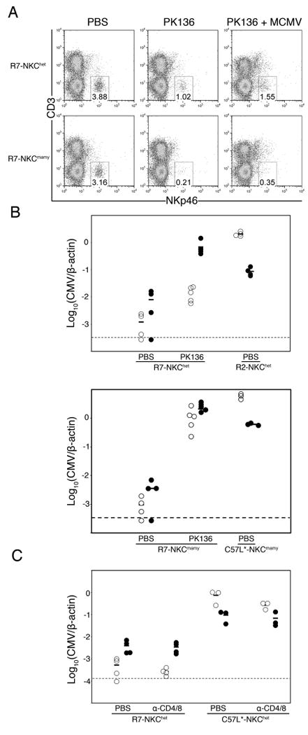
A. Splenocytes from R7-NKChet and R7-NKCmamy (Cmvr) mice given PBS, anti-NK1.1 mAb PK136, or PK136 followed by MCMV infection were stained for CD3 and NKp46. Numbers indicate the percentage of splenocytes within the NKp46+CD3- lymphocyte gate. B. R2 (Cmvs), R7 (Cmvr), C57L and PK136 treated mice were infected with MCMV. Shown are spleen (open) and liver (filled) MCMV genome levels for individual animals 90 h after infection. C. R7, C57L and T-cell-depleted (anti-CD4/CD8; see Materials and Methods) mice were infected with MCMV. Shown are spleen (open) and liver (filled) MCMV genome levels for individual animals 90 h after infection. Data included 3-5 mice per group.