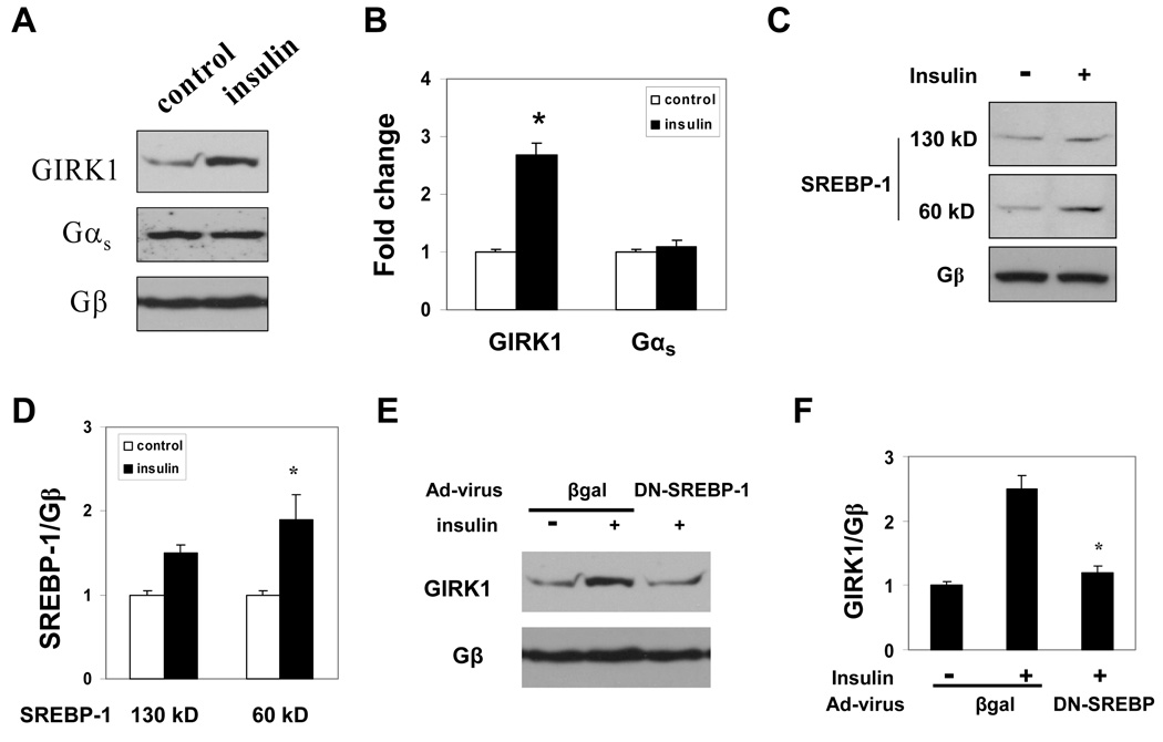
Figure 5. Insulin stimulation of GIRK1 expression in atrial myocytes is dependent on SREBP-1.
A. GIRK1 expression in embryonic chick atrial myocytes cultured for 16 hours in the presence and absence of 100 nM insulin determined by Western blot analysis. Only a single GIRK1 band was detected in the cultured atrial myocytes. Blots were sequentially stripped and reprobed with specific antibody to Gαs and Gβ. B. Mean intensity of GIRK1 determined by densitometry scanning of 3 independent experiments similar to that in A (*P<0.05). The relative intensity of GIRK1 normalized to Gβ in the absence of insulin was taken as 1. C. SREBP-1 levels in chick atrial myocytes cultured with and without insulin determined by Western blot analysis. D. Mean intensity of the precursor (130 kD) and processed (60 kD) SREBP-1 determined by densitometry scanning of 3 independent experiments similar to that in C (*P<0.05). E. Effect of DN-SREBP-1 on GIRK1 expression in cultured atrial myocytes. Embryonic chick atrial myocytes were infected with an adenoviral vector expressing GFP plus DN-SREBP-1 or GFP plus β-gal at the time of plating (MOI of 20). On the third culture day cells were incubated with or without insulin for 16 hours, harvested and expression of GIRK1 determined by Western blot analysis. F. Densitometry scanning of 3 experiments similar to that in E. Data are normalized to the expression of Gβ. *P<0.05.

