
Figure 1. Oncogenic v-rasHa does not block Smad phosphorylation or translocation.
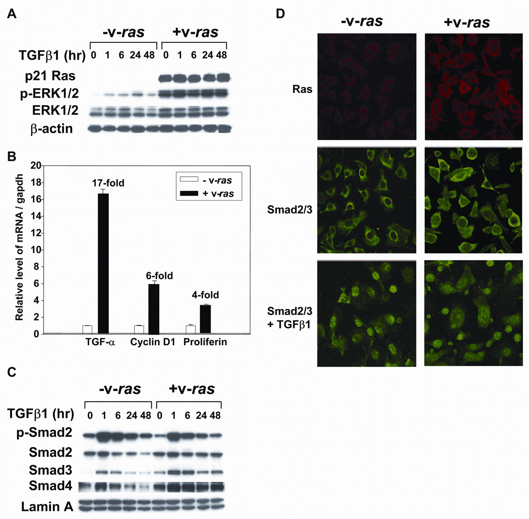
Balb/c primary keratinocytes were transduced with the v-rasHa retrovirus and then control and v-rasHa keratinocytes treated with TGFβ1 at 1ng/ml and samples taken for immunoblotting, quantitative PCR or immunofluorescence. A. Immunblotting of cellular extracts showing high levels of ras protein expression in transduced keratinocytes and elevated phosphorylation of ERK1/2 that is not altered by TGFβ1 treatment. B. Increased expression of proliferation associated genes in MEK expressing v-rasHa. RNA was isolated from control and v-rasHa transduced cells and expression of the indicated genes determined by quantitative PCR as described in Materials and Methods. The level of each transcript was normalized to that of Gapdh, and expressed as relative to the expression level in control keratinocytes. C. TGFβ1 induces similar alterations in Smad levels and phosphorylation in control and v-rasHa transduced keratinocytes. Nuclear extracts were isolated from keratinocytes at the indicated times after treatment with TGFβ1 and immunoblotted for the indicated Smads. D. v-rasHa does not prevent nuclear translocation of Smad2/3. Indirect immunofluorescence for Smad2/3 (FITC) or ras (Texas Red) in MEK or v-rasHa transduced MEK treated with TGFβ1 (1 ng/ml) for 1 hour. Nuclei were detected using DAPI staining.