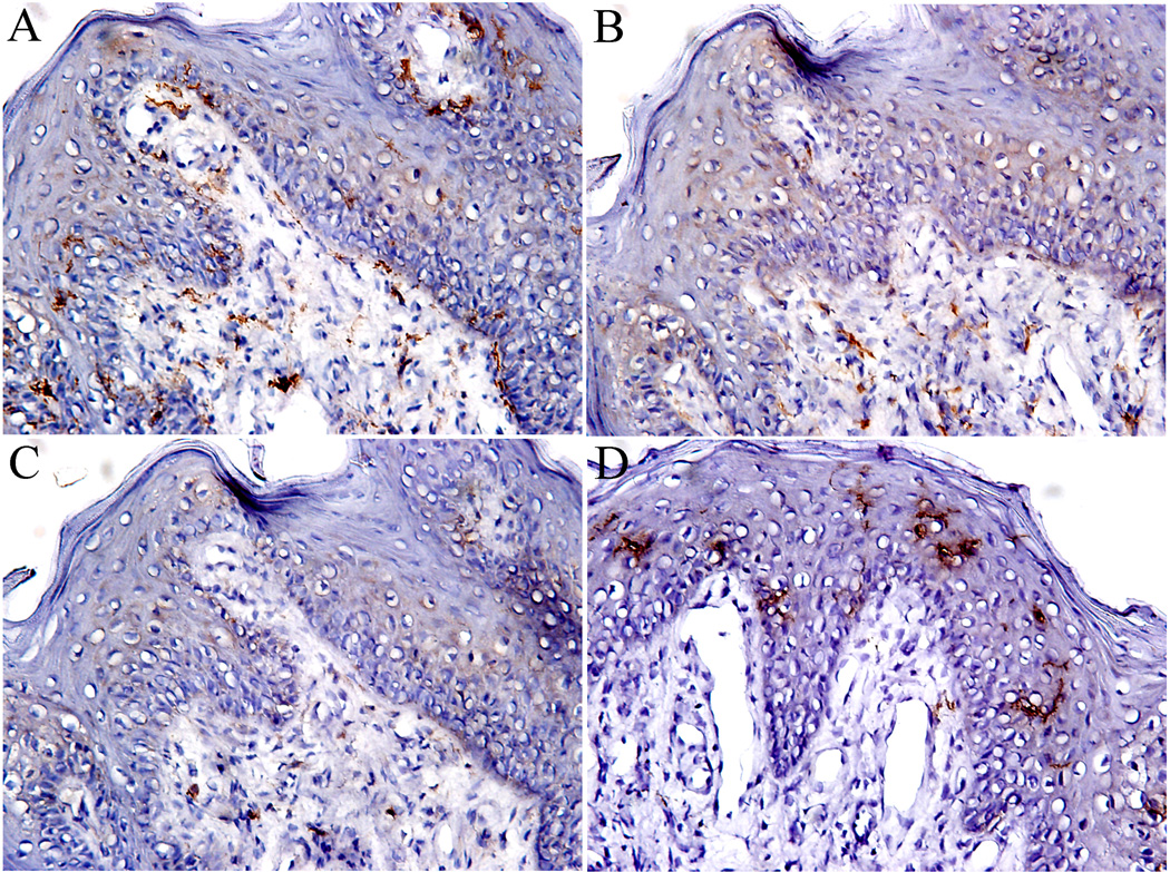
Figure 4.
Dendritic cells in fungiform papillae. A. CD11c staining dendritic cells; B. DC-SIGN staining immature dendritic cells; C. CD83 staining mature dendritic cells, and D. CD1a staining Langerhans cells. Note the difference in distributions of CD1a Langerhans cells, which are mainly present in epithelium, and CD11c dendritic cells, which are distributed both in epithelial and connective compartments. 10 × 20 magnification.

