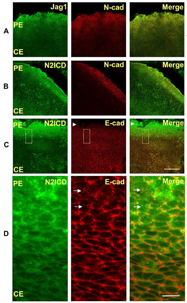
Fig. 1. Jag1 and N2ICD are highly expressed in the peripheral lens epithelium.
Representative immunofluorescence micrographs of whole mounts of freshly isolated postnatal day-2(PN2) rat lens epithelial explants stained with specific antibodies. (A,B) Jag1 (A) and N2ICD (B) expression colocalize with expression of N-cad, an early marker of differentiation, in the peripheral epithelium (PE). (C,D) The increase in the N2ICD corresponds to decreased immunostaining of Ecad (refer to arrowheads in panel C). Higher magnification (D), shown as an inset (dotted line box) in panel C, shows that the increased staining of N2ICD coincides with the appearance of intracellular vesicles (refer to arrows in panel D) that immunostain for E-cad, suggesting E-cad degradation (D). Scale bar on panel C represents 100 μm length and is applicable for all images of panels A, B and C. Scale bar on panel D represents 10 μm and is applicable for all images of panel D.

