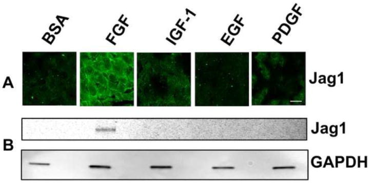
Fig. 5. Jag1 expression is not induced by IGF-1, EGF, or PDGF.
(A) Representative immunofluorescence micrographs of explants of central epithelium (CE) treated with BSA (control), FGF-2, IGF-1, EGF, or PDGF for 48 hours and immunostained for Jag1. Scale bar is applicable to all images of panel A and represents 10 μm length. (B) Corresponding immunoblots of lysates of treated explants from the same experiment immunoblotted for Jag1.

