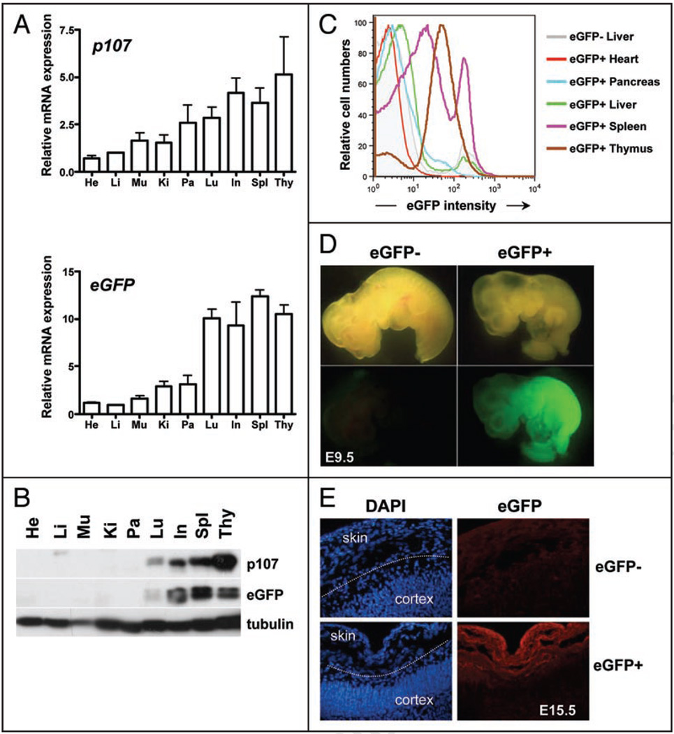
Figure 2.
Expression patterns of the eGFP transgene in adult mice and in embryos. (A) mRNA expression of p107 and eGFP relative to TBP (TATA Binding Protein) in whole organs from adult mice at 10–12 weeks of age (two mice—mean and standard error are graphed). (B) Representative immunoblot analysis of the p107 and eGFP proteins in extracts from whole adult mouse organs. He, heart; Li, liver; Mu, muscle; Ki, kidney; Pa, pancreas; Lu, lung; In, intestine; Spl, spleen; Thy, thymus. (C) Representative flow cytometric analysis of eGFP expression in single cell suspensions from whole organs obtained from adult p107 BAC-eGFP transgenic mice. The shaded grey area represents the eGFP signal for the liver of a non-transgenic mouse (eGFP−). Other organs are represented by lines of different colors, as indicated. (D) p107 BAC-eGFP is expressed throughout mid-gestation embryos (9.5 days of development, E9.5) by direct fluorescence. (E) Expression of the eGFP transgene in p107 BAC-eGFP mice becomes confined to proliferative regions such as the skin and not the maturing cortex in E15.5 embryos as demonstrated by indirect immunofluorescence using an anti-eGFP antibody (red)—DNA is visualized by DAPI staining (blue).

