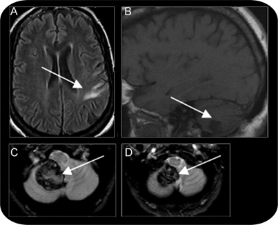
Figure MRI scans of the patient’s strokes
The foreign accent syndrome patient’s initial embolic middle cerebral artery stroke (A, axial image, fluid-attenuated inversion recovery sequence, lesion appears whiter than surrounding cortex) resulted in a lesion at the frontoparietal junction. The patient’s second stroke was a hemorrhagic stroke (3 cm × 4 cm × 1-3 cm hematoma) in the inferior portion of the right posterior lobe of the cerebellum reaching the midline and impinging on the vermis (T1-weighted sagittal image B, lesion appears darker than surrounding cortex, and susceptibility axial images C and D, where blood appears black). All images are presented in radiologic convention with the left hemisphere appearing on the right-hand side of the image. The white arrows indicate the lesioned area.

