
Figure 3.


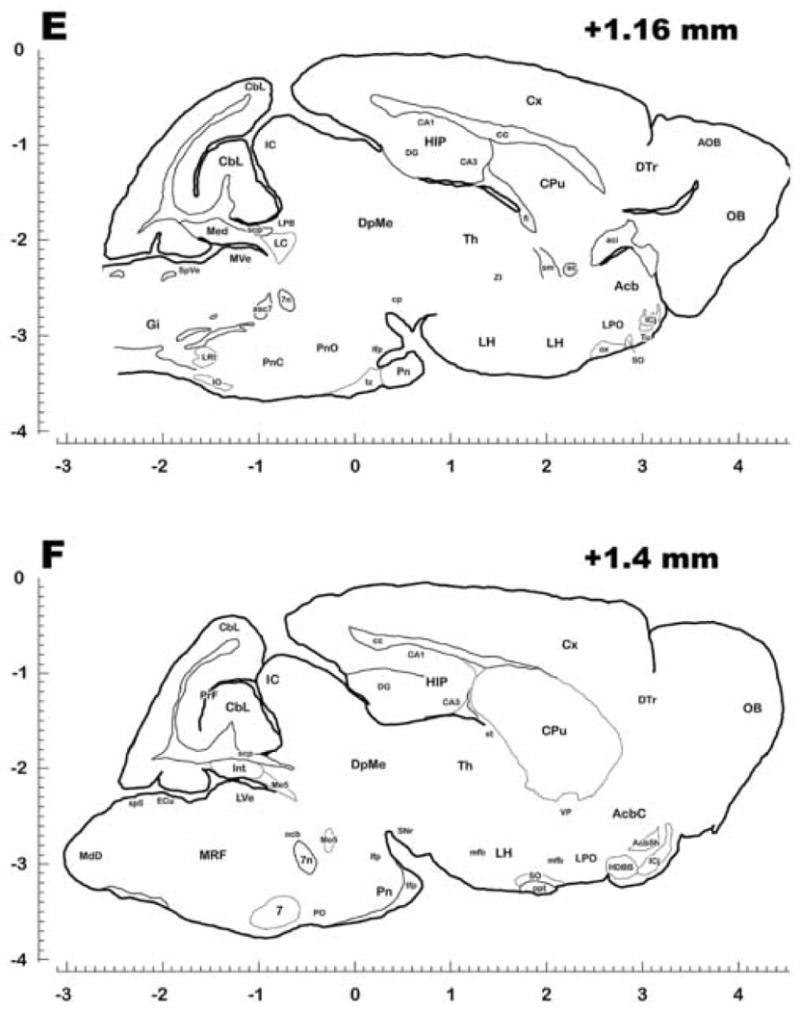

Representative Sagittal Sections of the Brain of Cryptotis Parva. Each section was traced from one or more complete, 40 μm thick sections stained with cresyl violet (Nissl) and/or Luxol Fast Blue (myelin), and embedded in gelatin to preserve the orientation and position of the various brain structures. Nuclei, and rarely subnuclei, were identified and labeled based on the atlases of Paxinos and Watson (rat atlas, 1998) and Slotnick (mouse atlas, 1975) respectively. Abbreviations conform to the conventions used in the Paxinos atlas and are also defined in Table 2. For stereotaxic measurements, scales are divided into 0.1 mm increments in the rostrocaudal and dorsoventral planes, with the value at the top right of each plate representing distance from the midline. Rostrocaudal zero was based on the apical junction of the lambdoid suture, and dorsoventral zero was based on the dorsal surface of the neocortex just adjacent to the midline. Plates proceed from medial (A) to lateral (H). The plates presented here are not meant to be a complete set, but are meant to represent as many major identified nuclei, fiber tracts, and key structures as possible without excessive redundancy.