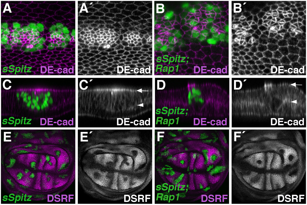
Figure 2. Rap1 and Egfr signaling affect different aspects of DE-cad localization.
(A–F) The MARCM system was used to express a secreted version of Spitz (sSpitz) in wildtype (A,C,E) or Rap1 mutant (B,D,F) cells. (A–D) Pupal wings (36 hours APF) stained for DE-cad are shown. (A) sSpitz expressing cells (GFP-positive) have high levels of DE-cad and are apically constricted compared to their wildtype neighbors. (B) Loss of Rap1 dramatically affects DE-cad localization in sSpitz expressing cells. (C,D) Optical cross-sections through the bi-layered wing epithelium are shown. Arrows and arrowheads indicate apical and basal surfaces of the dorsal wing epithelia respectively. (C) In sSpitz expressing cells, DE-cad is concentrated near the apical cell surface (apico-laterally). (D) Loss of Rap1 does not affect the apical/basal distribution of DE-cad in sSpitz expressing cells. (E,F) Wing imaginal discs stained for the intervein marker DSRF are shown. (E) sSpitz expressing cells downregulate DSRF indicating a switch to vein cell fate. (F) Loss of Rap1 does not affect the ability of sSpitz to downregulate DSRF.

