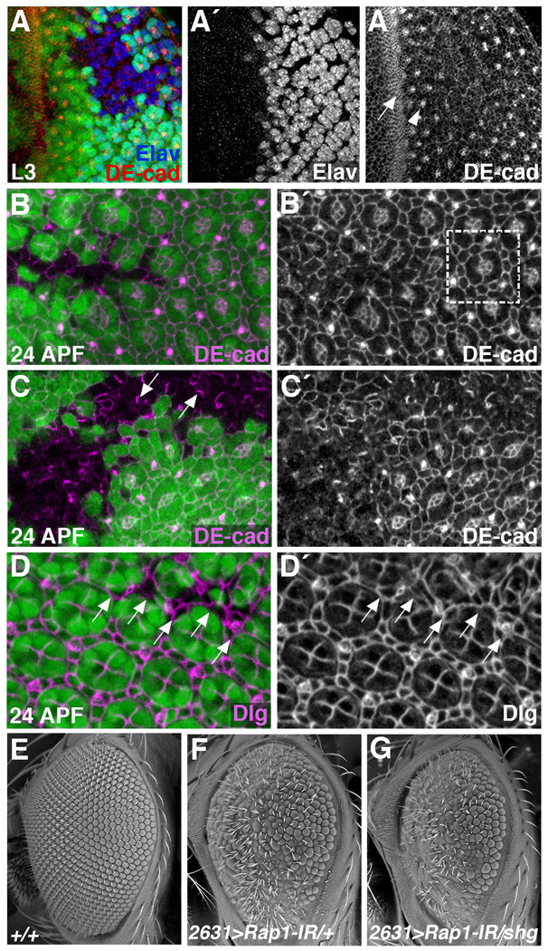
Figure 3. Rap1 affects cell adhesion and cell shape in the developing eye.
(A–D) Clones of cells lacking Rap1 (GFP negative) were generated using the Flp/FRT system. Eye tissue from late L3 larvae (A) or 24 hour APF pupae (B–) were stained for DE-cad (A–C) or Discs Large (Dlg) (D). (A) During larval stages, DE-cad levels are highest in the morphogenetic furrow (arrow) and at junctions between photoreceptor precursors (arrowhead). In Rap1 mutant cells, photoreceptors are still specified (A´), but the pattern of DE-cad localization is altered (A´´). (B,C) At 24 hours APF, DE-cad localization in the eye demarcates a regular lattice of cone and pigment cells. Boxed region in B´ indicates a single ommatidial unit. Loss of Rap1 disrupts DE-cad localization and the regular pattern of cell types. (C) Asymmetrical distributions of DE-cad are frequently observed in Rap1 mutant tissue (arrows). (D) Dlg localization similarly reveals a regular pattern of cone and pigment cells in wildtype 24 APF pupal eyes. Arrows point to Rap1 mutant cells, which are morphologically defective. (E–G) SEM images of adult eyes are shown. NP2631-Gal4, when crossed to a Rap1-RNAi transgene (Rap1-IR), knocks down Rap1 function in most eye cells. Compared to wildtype eyes (E), NP2631>Rap1-IR eyes are rough, typically indicating a defect in cell-type specification or differentiation (F). (G) The NP2631>Rap1-IR phenotype is significantly worsened when a single copy of the DE-cad/shg gene is eliminated.

