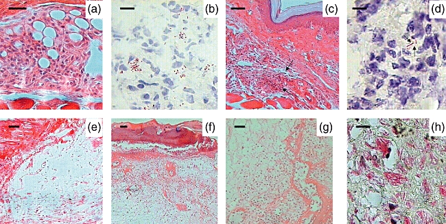
Fig. 3.

Representative histopathology of BALB/c subcutaneous footpad tissue infected with Mycobacterium ulcerans strains from Mexico and Benin. (a) A well-organized inflammatory infiltrate containing lymphocytes and macrophages is seen after 28 days of infection with strain 5114 from Mexico (400×, bar = 50 µ). (b) In the same lesion there are numerous extracellular bacilli as demonstrated by Ziehl–Neelsen staining (1000×, bar = 10 µ). (c) After 2 months of infection with M. ulcerans from Mexico, lymphocytes and macrophages are organized in small nodules forming granuloma-like structures (arrows) (40×, bar = 200 µ). (d) In these lesions, occasional bacilli were found (arrows) (1000×, bar = 10 µ). (e) Extensive oedema with slight inflammatory infiltrate is seen after 1 month of infection with strain 1441 from Benin (100×, bar = 100 µ). (f) After 2 months of infection, the Benin strain induced extensive oedema, massive necrosis with acantholysis and a slight inflammatory infiltrate (40×, bar = 200 µ). (g) In the same section, the connective tissue around the necrotic areas shows abundant chronic inflammatory infiltrate (40×, bar = 200 µ). (h) These necrotic areas show massive amount of extracellular bacilli revealed by Ziehl–Neelsen staining (1000×, bar = 10 µ).
