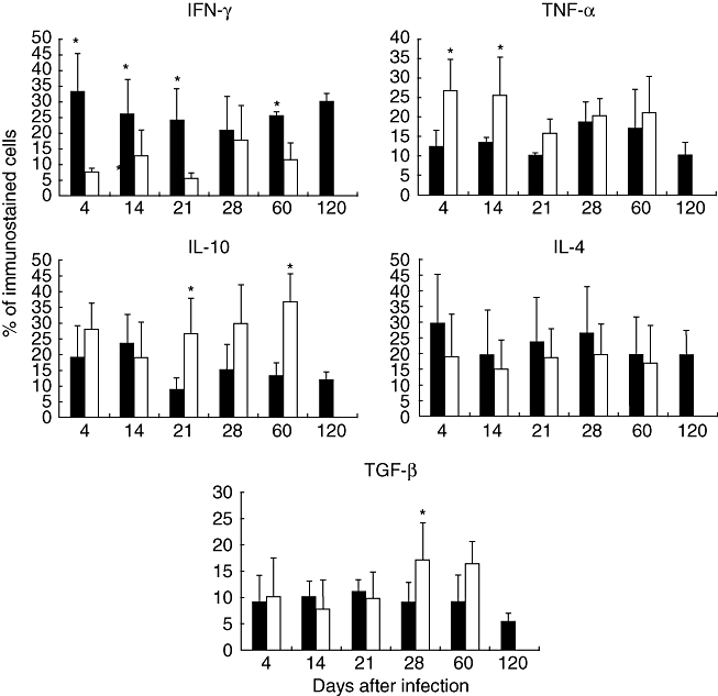
Fig. 5.

Determination of percentages of cells immunostained for cytokine production during infection with Mycobacterium ulcerans from Mexico (black bars) and Benin (white bars). Three different randomly chosen areas were used to determine, by immunohistochemistry and automated morphometry, the number of positive and negative cells in the inflammatory infiltrate of three different mice per time-point. Then, the percentage of positive cells was determined for each cytokine indicated. Data are expressed as mean and standard deviation; asterisks represent statistical significance (P < 0·005).
