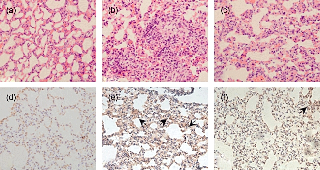
Fig. 4.

Haematoxylin and eosin staining and immunohistochemistry staining of the lung tissues in mice (×100). (a–c) Haematoxylin and eosin staining; (d–f) immunohistochemistry staining of α-smooth muscle actin (α-SMA). (a,d) Group n; (b,e) group bleomycin (BLM); (c,f) group thalidomide (THD). Group n: mice injected with phosphate-buffered saline subcutaneously and gastric perfused with carboxymethyl cellulose (CMC); group BLM: mice injected with BLM subcutaneously and gastric perfused with CMC; group THD: mice injected with BLM subcutaneously and gastric perfused with THD.
