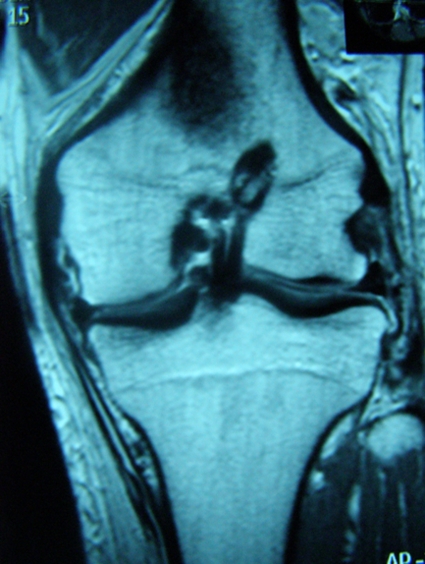
Fig. 1-A Fig. 1-A.
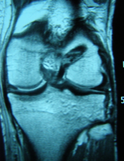
Figs. 1-A and 1-B Postoperative magnetic resonance imaging scans showing the two different positions for the femoral tunnel used in this study. Fig. 1-A The femoral tunnel was drilled at the one o'clock position (corresponding to the eleven o'clock position in a right knee). Fig. 1-B The femoral tunnel was drilled more laterally, at the two o'clock position (corresponding to the ten o'clock position in a right knee).


