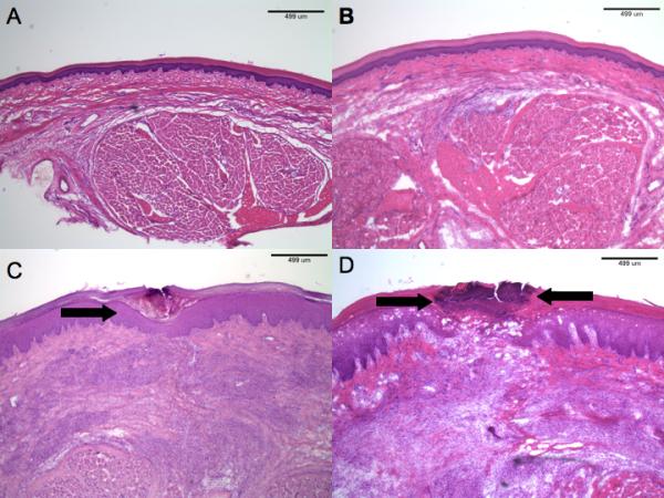
Fig. 6.

(A) H & E section of normal rat hindpaw. (B) Low-power histopathology of sham-treated hindpaw after (50μg/200μl) was injected. There are no abnormalities. (H & E). (C) Histopathology of incised rats receiving vehicle. Note the superficial scale-crust (arrow), but otherwise normal-appearing epidermis. The dermis shows granulation tissue around the site of incision. (H & E). (D) Incised hindpaw histopathology in a rat receiving subcutaneous capsaicin injection (200μg/200μl). There is pronounced, full-thickness, epidermal necrosis (region between arrows). The dermal findings reveal granulation tissue similar to that observed in vehicle-only treated rats. (H & E).
