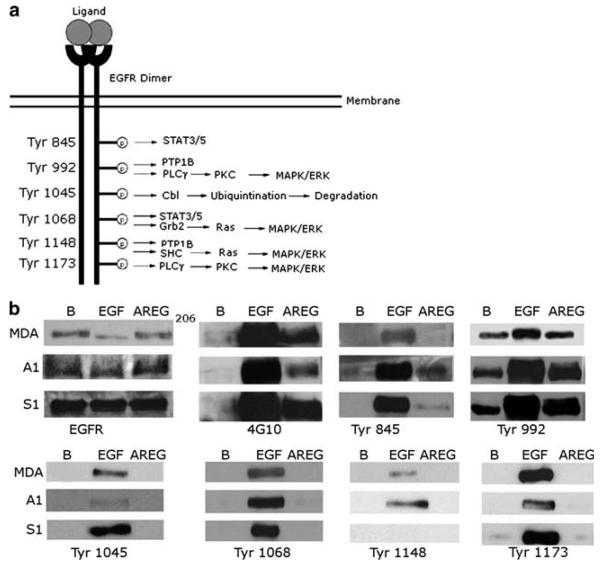
Fig. 3.
AREG stimulates a distinct pattern of EGFR tyrosine phosphorylation. The A1, S1 and MDA-MB-231 cells were stimulated with 10 nM of EGF, 100 nM of AREG, or PBS (B). Glycoproteins from the protein extracts were precipitated with concanavalin A sepharose and resolved by SDS-PAGE. The immunoblots were then probed with the corresponding antibody. This experiment was repeated three times

