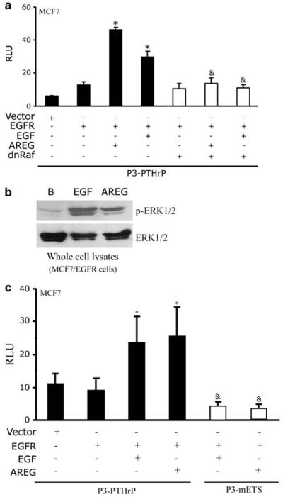
Fig. 8.
Identification of signaling components downstream of EGFR that activate PTHrP gene expression. (a) and (c) Transient transfections assay comparing the reporter activity of the wild-type EGFR (WT), EGF (EGF) or AREG (AREG)-stimulated wild-type EGFR (WT) in the presence or absence of dnRaf or with a P3-mETS reporter. The luciferase activities were normalized to β-galactosidase levels and were expressed as means ± s.d. of triplicate cultures. RLU, relative luciferase units. (b) MCF7/EGFR cells were treated for 10 min with 10 nM EGF or 100 nM AREG. Whole cell lysates were harvested and resolved by SDS-PAGE. The immunoblots were then probed with the corresponding antibody. This experiment was repeated twice. *P < 0.01 vs. unstimulated EGFR &P < 0.01 vs. AREG or EGF stimulated EGFR

