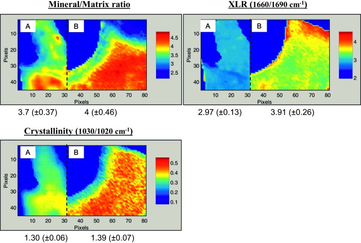
FIG. 1.
Typical infrared images for the FTIR parameters recorded in trabecular bone from two patients, one with fractures and one without, who had comparable BMD T-scores of −1.3. Patient A had no fractures (t = −1.32) and was 50 yr old at time of biopsy. Patient B had a fracture history (T = −1.25) and was 58 yr old at time of biopsy. Numerical values below the images are the means ± SD for that parameter in the figure and indicate the range of data for the pixels shown. Note in these figures, 1 pixel = 6.25 μm.

