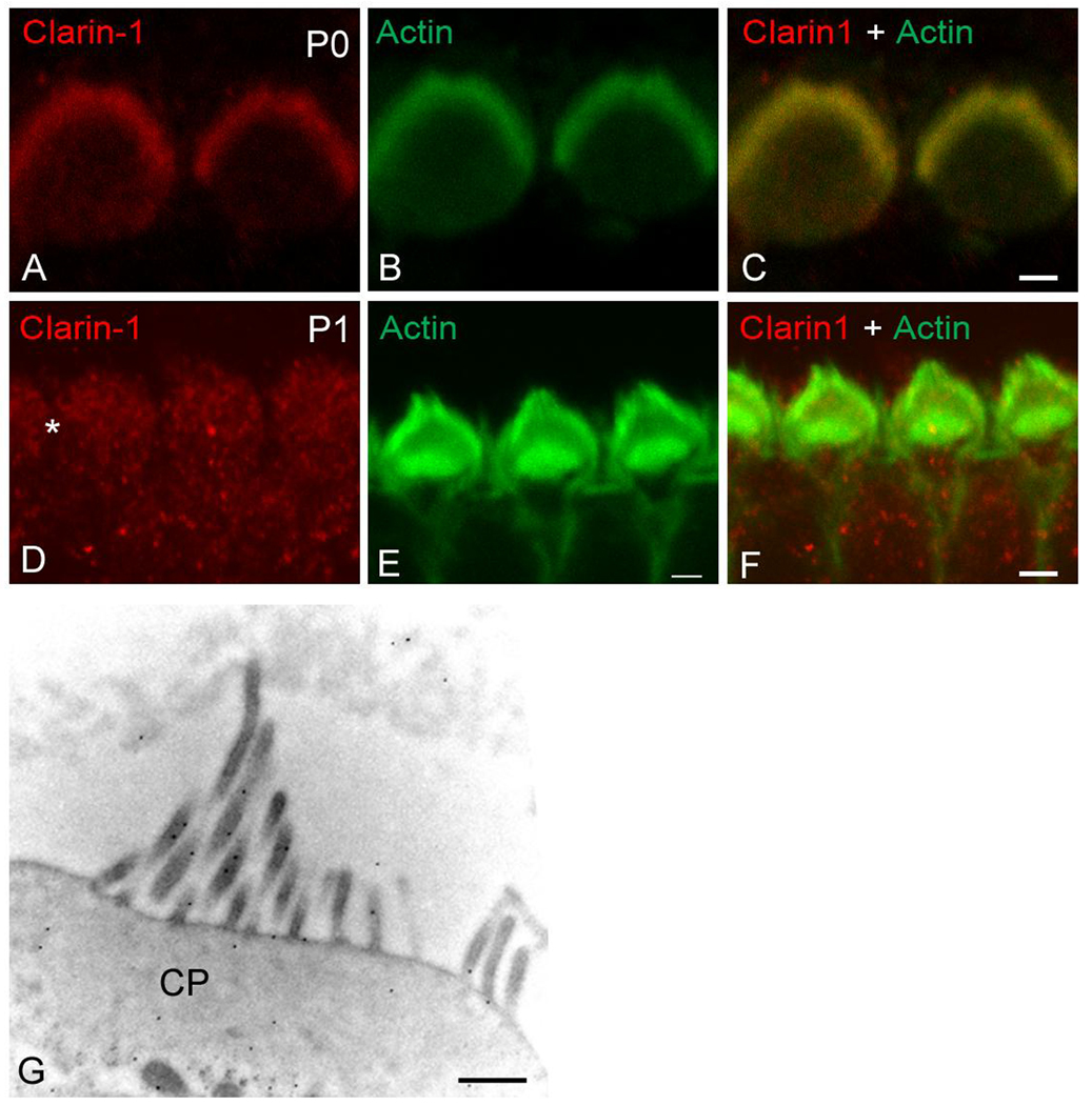
Figure 4.
Expression of clarin-1 at the apical aspect of hair cells. Surface preparations of P0 (A–C) and P1 (D–F) organs of Corti were analyzed by confocal microscopy. A, D: immunostained for clarin-1. B, E: counterstained with phalloidin. C, F: merge image. Asterisk denotes weak immunostaining for clarin-1 in the cell bodies. G: Immunogold localization of clarin-1 in outer hair cells from P0 mice. Ultrastructural localization is consistent with low resolution localization shown in Figure 3. CP: cuticular plate. Scale bar: A–C: 1 µm, D–F: 2 µm, G: 500 nm.

